Premature and Early Menopause: Symptoms and Treatments

Is Hypnosis Real and How Does It Work? What Does Science Say?
Table of Contents
- What is Hypnosis and How Does It Work on the Brain?
- The Science Behind How Hypnosis Works on the Brain
- What Happens in the Brain during Hypnosis?
- Anatomy of a Clinical Hypnosis Session
- What Does Hypnosis Feel Like in the Brain?
- What Are the Signs That You Were Hypnotized?
- How Long Can the Effects of Hypnosis Last on the Brain?
- How to Use Self-Hypnosis to Influence the Brain
- Tips for Making Self-Hypnosis Effective on Your Brain
How could you make better use of your brain? For example, did you know you could train your brain to let go of pain and negative habits like smoking? Science has started to uncover how hypnosis works on the brain. And the findings are truly fascinating.
This blog will provide an in-depth exploration of how hypnosis works on the brain, its effects, and how to use self-hypnosis to improve your life. It will cover topics such as the effects of hypnosis on the brain and what happens when you get hypnotized. We will also review the signs that you were hypnotized and explain how long you can stay hypnotized.
What is Hypnosis and How Does It Work on the Brain?
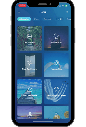
Hypnosis represents both a state of focused attention and the techniques used to create this state. The utilization of hypnotic methods for therapeutic purposes is called hypnotherapy. As science became better at understanding the brain, doctors and therapists have become aware of its clinical potential and started incorporating hypnosis in their practice. Today, you can enjoy the benefits of hypnotherapy from home through a self-hypnosis app.
The Science Behind How Hypnosis Works on the Brain
Scientists have researched hypnosis for centuries. Since Mesmer became first interested in this field, scientists went through cycles of intense curiosity followed by complete disinterest. More noticeably, this wave of modern exploration started in Europe with Mesmer and then Dr Liebault and Dr Charcot.
The last two doctors were French and studied hypnosis in the second half of the 19th century with the means at their disposal. Dr Jean-Martin Charcot (1825-1893) was a neurologist and Dr Ambroise-Auguste Liébeault (1823-1904) a physician.
Liébeault believed that hypnosis could be used to treat many psychological issues and medical conditions, and he used it extensively in his clinical practice. He also trained many other physicians and therapists in the use of hypnosis, and his approach to hypnotherapy became known as the Nancy School of Hypnosis.
Liebault set up a clinic visited by many French and European doctors who were, like Sigmund Freud, interested in hypnotism. Liébeault’s work was instrumental in shaping the early development of hypnotherapy as a clinical practice, and his ideas continue to influence the field today. However, his work was largely overshadowed by the more famous figures in the history of hypnosis, such as James Braid and Sigmund Freud, and he is not yet as well-known outside of the field of hypnotherapy.
Dr Charcot opened his own clinic and while his focus was on neurology, the two clinics competed for thought leadership and by trying to outdo each other contributed to great advances in understanding the field of clinical hypnosis. It was the first time that Western medicine would consider the brain-body connection. We consider these scientists the first pioneers of clinical and experimental hypnosis as a tool to treat mind and body.
What Happens in the Brain during Hypnosis?
The latest developments in brain imaging studies shed light on the fascinating effects of hypnosis on the brain. Technology is now meeting an old practice already utilized by therapists and doctors for generations, helping us understand how hypnosis works on the brain. Before reviewing some of the studies, let’s look at the modern tools used in hypnosis research.
Different Brain Imaging Techniques Used in Research
Medical research has used many types of brain imaging techniques to study the effects of hypnosis on the brain. These tools have truly advanced our knowledge in cognitive and clinical neuroscience. They measure magnetic fields, and electrical currents on the scalp or track the distribution of a particular molecule to gain a deeper understanding of the anatomy, metabolism, and activity of the brain. Brain scanning can also study functional connectivity between brain areas.
These tools include electroencephalography (i.e EEG), positron emission photography, functional magnetic resonance imaging, magnetoencephalography, and computed tomography. We will focus on the first three techniques.
EEGs: the First Tool to Study Brain Regions
First developed in 1920s, electroencephalography uses electrodes placed on the scalp to measure the brain’s electrical activity. With this technique, researchers can study brain function and activity, including the rhythms and patterns of neural activity associated with different mental states and of consciousness.
Positron Emission Tomography (PET)
Positron emission tomography (PET) uses a radioactive tracer to track the distribution of a specific molecule in the brain, such as glucose or dopamine.
The successful mapping of these molecules’ journeys can enhance our comprehension of brain function and metabolism, particularly when studying neurological and psychiatric disorders. This technique was developed in the 1970s.
Functional Magnetic Resonance Imaging
Functional magnetic resonance imaging (or fMRI) is one of the most recently created method. Developed in the 1990s, fMRI uses strong magnetic fields and radio signals to generate detailed images of the brain’s structure.
By measuring changes in blood flow in different brain areas, fMRI can help track changes in neural activity. This technique can be used to study brain function, including the areas of the brain that are activated during various tasks or in response to different stimuli.
What Part of The Brain Is Affected By Hypnosis?
Brain Wave Patterns
Because the brainwave frequency slows down and reaches the theta band (from 4 to 7Hz) in hypnosis, you could argue that the whole brain is affected during this trance-like state. A person able to enjoy deep relaxation, daydreaming and meditation also sees their brain function differently.
Scientists uncovered the transition from one state to another based on the brainwaves’ variations with EEGs. The only challenge with EEGs is the absence of a spatial map. You cannot know where the electrical signal originated from.
fMRI and other imaging techniques helped open the doors to understanding the effects of hypnosis on the brain. Let’s review the most notable findings.
Decreased Activity In Default Mode Network
The default mode network (i.e. DMN) is activated when a person is either thinking, remembering, daydreaming and not carrying out a task. If a person starts to focus on a goal or becomes absorbed by something, the DMN is inactive. Said differently, a decrease in DMN activity leads to a greater sense of absorption and attention, also called attentional absorption.
Researchers noted a reduction in activity in the brain’s default mode network in those undergoing hypnosis, supporting the definition of hypnosis as a state of focused attention.
Increased activity in the Prefrontal Cortex
The prefrontal cortex, located in the brain’s frontal areas, is responsible for executive functions like problem-solving, reasoning, and planning. Research shows that hypnosis can significantly increase activity in the prefrontal cortex, enhancing a person’s ability to focus and concentrate. This finding demonstrates how hypnosis works on the brain, particularly in areas related to cognitive control.
Reduced activation in the Dorsal Anterior Cingulate Cortex
The dorsolateral prefrontal cortex is a critical part of the executive control system, while the posterior cingulate cortex is associated with self-related thinking. The effects of hypnosis on the brain include reduced connectivity between these areas, which may explain the dissociative experiences common during hypnosis.
Modulated activity in brains areas involved in the regulation of consciousness
PET scans have shown that hypnosis increases blood flow in brain regions involved in consciousness regulation, such as the anterior cingulate cortex (ACC), thalamus, and brainstem. These findings underscore how hypnosis works on the brain by modulating areas responsible for consciousness and self-awareness.

Reduced Connectivity Between the Dorsolateral Prefrontal Cortex and the Posterior Cingulate Cortex
The effects of hypnosis on the brain extend to reduced connectivity between the dorsolateral prefrontal cortex (involved in executive control) and the posterior cingulate cortex (part of the default mode network). This reduced connectivity is thought to play a role in the dissociative effects experienced during hypnosis.
Increased Connectivity Between the Insula and the Dorsolateral Prefrontal Cortex
The insula is brain area located deep within the cerebral cortex. The insula lobe is involved in a variety of cognitive and emotional processes, including self-awareness, perception, emotion processing, motor control and autonomic functions. As a result, the insula plays an important role in decision-making, pain processing and reward anticipation.
This increased connectivity could explain why a person in hypnosis has greater control over physical symptoms. They can change their pain perception and temperature. To this date, pain control is one of the most used applications in clinical settings. Other autonomic functions that can be controlled under hypnosis are heart rate, blood pressure, respiration and digestion.
Some studies have also suggested that hypnosis can also be utilized to reduce symptoms of anxiety by inducing relaxation and reducing physiological arousal.
What Part of the Brain does Hypnosis Activate?
How Does Hypnosis Work in the Brain? Because the brain is complex, this simple question calls for a complex response.
Medical imaging unearthed many hypnosis effects on the brain. For example, hypnosis causes changes in the brain connectivity and activity consistent with greater control on internal sensations and emotions, reduced fear and reduced self-consciousness.
Myths and Misconceptions
Myths and misconceptions about hypnosis are fading as more psychotherapists and doctors educate patients on hypnosis. Let’s summarize the most common.
- Researchers are categorical. Hypnosis is not sleep. Brainwaves during hypnotic trances are not sleep brainwaves.
- You do not lose control in hypnosis. You regain control over your body and you can improve your mental health.
- You cannot be made to do anything you don’t want to do. Did you notice that a hypnotist keeps sending people back to their seats in a stage hypnosis show because they are not compliant? And all these movements are done under the guise of “exercises”?
Anatomy of a Clinical Hypnosis Session
A hypnotherapist helps a person revert to the hypnotic state through a hypnotic induction, followed by some deepening work. When you reach this heightened state of suggestibility, the hypnotherapist guides you through hypnotic suggestions crafted to help you achieve deep sleep at night, or enjoy greater physical relaxation. The therapist then helps you emerge and concludes the hypnotherapy session with some more suggestions.
What Does Hypnosis Feel Like in the Brain?
If you asked participants in the same hypnosis session to describe their experience, you would hear that it was very relaxing and they remained aware of what was happening but then you would receive many different depictions of this trance-like state.
Some people typically describe feelings of lightness, some have feelings of heaviness and the rest do not feel much difference in the body. Everyone has a unique experience.
What Are the Signs That You Were Hypnotized?
How do you recognize a hypnotic state? Remember that with a hypnotherapist, you learn to enter self-hypnosis. You are far from passive: you are not hypnotized; you are recreating this state of deep focus. Here are many ways to notice whether you are in a hypnotic state:
- more relaxation and focus
- changes in your breathing and heart rate
- altered sensations such as heaviness, tingling, or numbness
- vivid imagery
- sense of detachment from time and space
- reduced anxiety levels
- increased confidence and improved mental clarity.
- and whichever clinically proven goal you are working on.
How Long Can the Effects of Hypnosis Last on the Brain?
The length of time that a person can remain in a hypnotic state varies from individual to individual and from session to session. Generally speaking, the average length of time for a successful hypnosis session is between 10 to 30 minutes. Some people may be able to remain in an extended state of hypnosis for up to an hour or more, depending on the particular circumstances.
How to Use Self-Hypnosis to Influence the Brain
Self-hypnosis is not a magic cure. It requires practice. Brain plasticity is a wonderful thing because our nervous system continues to evolve but you need to repeat a movement or a particular thought pattern a few times to make it second nature. A self-hypnosis app helps you do just that: enjoy sessions 10 to 30 minutes long where you can revel in deep focused attention leading to a reverie state.
Benefits of Clinical Hypnosis
Many medical practitioners, psychotherapists and professional hypnotherapists use hypnosis for therapeutic purposes:
- quitting smoking
- reducing anxiety and stress
- treating pain
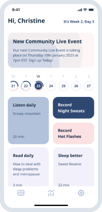
- managing menopause (reducing hot flashes and night sweats)
- achieving peak performance
- managing IBS symptoms
- increasing self-confidence and self-esteem
- lose weight
Tips for Making Self-Hypnosis Effective on Your Brain
Carve out some time in your calendar and make sure to switch off any distractions. Put on your headset and start your daily hypnosis audio. Allow yourself to relax deeply and listen to your hypnotherapist’s guided suggestions. During these few minutes of hypnosis, you can forget about everything else and enjoy this deep state of focus. When you emerge, you can feel more energized and ready to go with your day!
Hypnosis has many applications, from treating smoking addiction to managing chronic pain or even just feeling good. More research continues to uncover more benefits of this altered state of consciousness. Download UpNow hypnosis today! Get started now.
UpNow Health only uses high-quality sources, including peer-reviewed articles, to support the facts within our articles. Experts review all our articles to ensure that our content is accurate, helpful, and trustworthy.
1. Rainville, P., & Price, D. D. (2003). Hypnosis phenomenology & the neurobiology of consciousness. The International journal of clinical and experimental hypnosis, 51(2), 105–129. https://doi.org/10.1076/iceh.51.2.105.14613
2. Rainville, P., Hofbauer, R. K., Bushnell, M. C., Duncan, G. H., & Price, D. D. (2002). Hypnosis modulates activity in brain structures involved in the regulation of consciousness. Journal of cognitive neuroscience, 14(6), 887–901. https://doi.org/10.1162/089892902760191117
3. Jiang, H., White, M. S., Greicius, M. D., Waelde, L. C., & Spiegel, D. (2016). Brain Activity and Functional Connectivity Associated with Hypnosis. Cerebral Cortex. https://doi.org/10.1093/cercor/bhw220
4. Rainville, P., Hofbauer, R. K., Bushnell, M. C., Duncan, G. H., & Price, D. D. (2002). Hypnosis modulates activity in brain structures involved in the regulation of consciousness. Journal of cognitive neuroscience, 14(6), 887–901. https://doi.org/10.1162/089892902760191117
5. Müller, K., Bacht, K., Prochnow, D., Schramm, S., & Seitz, R. J. (2013). Activation of thalamus in motor imagery results from gating by hypnosis. NeuroImage, 66, 361–367. https://doi.org/10.1016/j.neuroimage.2012.10.073
6. Raij, T. T., Numminen, J., Närvänen, S., Hiltunen, J., & Hari, R. (2009). Strength of prefrontal activation predicts intensity of suggestion-induced pain. Human brain mapping, 30(9), 2890–2897. https://doi.org/10.1002/hbm.20716
7. Cojan, Y., Waber, L., Schwartz, S., Rossier, L., Forster, A., & Vuilleumier, P. (2009). The brain under self-control: modulation of inhibitory and monitoring cortical networks during hypnotic paralysis. Neuron, 62(6), 862–875. https://doi.org/10.1016/j.neuron.2009.05.021
8. Vanhaudenhuyse, A., Boly, M., Balteau, E., Schnakers, C., Moonen, G., Luxen, A., Lamy, M., Degueldre, C., Brichant, J. F., Maquet, P., Laureys, S., & Faymonville, M. E. (2009). Pain and non-pain processing during hypnosis: a thulium-YAG event-related fMRI study. NeuroImage, 47(3), 1047–1054. https://doi.org/10.1016/j.neuroimage.2009.05.031
9. Deeley, Q., Oakley, D. A., Toone, B., Giampietro, V., Brammer, M. J., Williams, S. C., & Halligan, P. W. (2012). Modulating the default mode network using hypnosis. The International journal of clinical and experimental hypnosis, 60(2), 206–228. https://doi.org/10.1080/00207144.2012.648070


